
This section assumes knowledge from the sections: DNA, Basic biology, Cancer, Mitosis and meiosis.
Cancer diagnosis is a complex task that takes years of medical training to properly understand and administer, this section is not a guide for cancer diagnosis but a summary of actual tests medical staff can do that may be confusing, intimidating or interesting.
There are 2 main types of diagnostic tests that are not intuitive to most and thus will be covered in this section. The first are scans.
Scans
CT scan (computed tomography): this is a device that produces X-ray radiation and spins around the patient. As the radiation passes through the patient’s body its energy will emerge on the other side differently depending on the tissue the radiation passes through. Reading the differences in this energy can allow the computer to generate an image of a cross section of the person’s body. Trained medical professionals know what healthy scans look like and can identify oddities in the images that may be tumors.
(1)
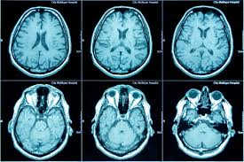
Fig 1: CT scan images of a person’s brain.
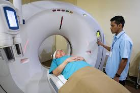
Fig 2 : Image of a person positioned inside a CT scanner
MRI (magnetic resonance imaging): MRI machines use magnetic fields to align the protons within a person’s body. The machine can then introduce a radiofrequency pulse to briefly realign the protons. The speed at which different areas then return to the magnetic field indicates what type of tissue it is, because different tissues have different amounts and structures of protons. (2)
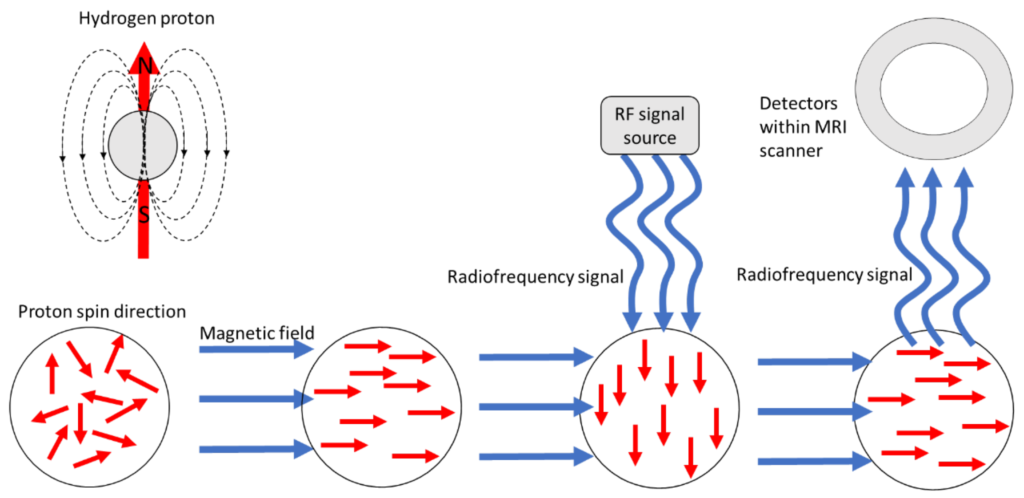
Fig 3 : A diagram showing how MRI machines affect protons.
Nuclear scan: Nuclear scans are an umbrella term that incorporates a variety of scans(a well known nuclear scan is a PET scan), however they all function under the same principle and will therefore be grouped into a singular section. Nuclear scans work by introducing a radioactive material called a tracer into the body, technology outside of the body can then pick up the radiation coming from the tracer and form an image of where it is. This image can be used to evaluate a number of things and is especially useful for measuring how the pathways within the body are functioning. Fig 4 is an image of a lung that has had a nuclear scan, as you can see in the second set of lungs certain areas have less blood flow and have therefore received less of the tracer.
(3)
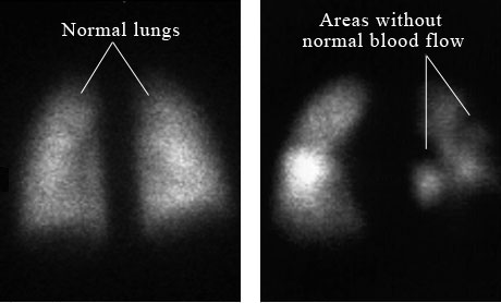
Fig 4: Results from nuclear scans of 2 peoples lungs
Ultrasounds: ultrasounds are a scan using high frequency sound waves, they are popular for uses in prenatal care as it is non-invasive and does not use radiation, however it can still be used to find abnormal physical formations in the body such as tumors. Sound waves will emit from the device and depending on the tissues it passes through different soundwaves will return allowing computers to build an image. It could be compared to echolocation, using soundwaves to map out physical objects based on how they come back.
(4)
X-rays: X-rays are also their own class of scans, X-rays are usually used to easily view bones, cavities and foreign objects. They differ from CT scans because they function on a 2D plan, firing X-ray radiation from 1 point to create an image.
Tests done on samples
The second form of diagnostic test to cover are lab tests done on samples. The three most common ways of collecting samples are, firstly, through surgery where surgeons cut out small sections of tissue to be tested. Secondly, with needles, where needles are used to puncture skin and suck up a sample. And thirdly, endoscopy where a flexible tube often with a flashlight and camera are inserted into the body to take small samples from the digestive tract.
Note: endoscopy can be considered a type of surgery.
Cbc: Complete blood count is a counting of the cells within blood samples. A manual way of doing that is to put them into a counting chamber, diluting the solution and using a microscope to observe the correct amount of cells within a set area. This can inform medical staff if the body is producing the correct amount of each blood cell type.
(5)
Blood chemical tests: tests on the plasma of the blood, not the plasma cells but the plasma which is the liquid component of our blood. This is usually used to give a guide as to whether organs related to blood are functioning correctly, or to check for chemicals that may be produced by or in response to cancer.
(6)
Cytogenetic analysis: the analysis of chromosomes/DNA. There are a number of ways this can be done in relation to cancer. It is possible to dye the chromosomes and witness them during metaphase through a light microscope, this is called karyotyping. Comparative genomic hybridisation disassembles the chromosomes of a healthy regular cell and tumor cell, then attaches fluorescent proteins to them, then it is possible to compare the difference in genetic material between the two in specific chromosomes quantifying the loss or gain of genetic material by the fluorescence. Another type is array comparative genomic hybridization, it is similar however it uses probes to bind to the fluorescent DNA and assess smaller changes in the amount of genetic material.
(7)
Immunophenotyping: in immunophenotyping medical staff will create unique antibodies that have been coupled with a fluorescent component, by documenting which protein each antibody targets and which color those antibodies have it is possible to then document amounts and positions of proteins within samples. This can test for specific proteins that are produced by or in response to cancer, or to check cancer cells for differences in proteins that could be used to treat or classify it.
(8)
Cytological analysis: cytology is simply the study of individual cells and by analyzing various samples under a microscope for changes in normal cell structure it is possible to determine mutations. This practice can sometimes be referred to by the specific sample being analyzed such as urine cytology or sputum cytology.
(9)
(10)
| 1. | NIH. Computed Tomography (CT). National Institute of Biomedical Imaging and Bioengineering. Published 2022. Accessed July 11, 2026. https://www.nibib.nih.gov/science-education/science-topics/computed-tomography-ct |
| 2. | National Institute of Biomedical imaging and Bioengineering. Magnetic Resonance Imaging (MRI). National Institute of Biomedical Imaging and Bioengineering. Published 2025. Accessed July 11, 2026. https://www.nibib.nih.gov/science-education/science-topics/magnetic-resonance-imaging-mri |
| 3. | Cleveland Clinic. Nuclear Medicine Imaging. Cleveland Clinic. Published April 20, 2021. Accessed July 11, 2026. https://my.clevelandclinic.org/health/diagnostics/4902-nuclear-medicine-imaging |
| 4. | Health Direct. Ultrasound. Healthdirect.gov.au. Published January 29, 2019. Accessed July 11, 2026. https://www.healthdirect.gov.au/ultrasound |
| 5. | badr. 3..Manual CBC.pptx. Slideshare. Published October 22, 2022. Accessed July 11, 2026. https://www.slideshare.net/slideshow/3manual-cbcpptx/253760390 |
| 6. | Lee S. Blood chemistry tests. Canadian Cancer Society. Published 2019. Accessed July 11, 2026. https://cancer.ca/en/treatments/tests-and-procedures/blood-chemistry-tests |
| 7. | Smith E. Cytogenetic testing | DermNet NZ. dermnetnz.org. Published April 2019. Accessed July 11, 2026. https://dermnetnz.org/topics/cytogenetic-testing |
| 8. | Herold NC, Mitra P. Immunophenotyping. PubMed. Published 2020. Accessed July 11, 2026. https://www.ncbi.nlm.nih.gov/books/NBK558927/ |
| 9. | Urine Cytology: What Is It, Purpose, Procedure & Results. Cleveland Clinic. Published May 5, 2022. Accessed July 11, 2026. https://my.clevelandclinic.org/health/diagnostics/22942-urine-cytology |
| 10. | Shen F, Sergi C. Sputum analysis. PubMed. Published 2023. Accessed July 11, 2026. https://www.ncbi.nlm.nih.gov/books/NBK563195/ |