
Another key process to do with cancer is mitosis. Mitosis is the process cells use in order to replicate themselves. This is how multicellular organisms (such as humans) grow and repair damage, and replace older cells. Mitosis is a well documented and easily observed process, with a regular light microscope and the correct dyes it is possible to watch mitosis in action. This concept can be explored in as little or as much depth as a reader would like. Conceptually mitosis is a very simple process, the cell will make duplicates of its organelles, it will then begin to expand, and eventually it will constrict and separate into 2 separate cells that are identical to the original cells with their own membranes.
To understand treatments and intricacies of cancer it is necessary to understand growth factors, the cell cycle and the stages of mitosis.
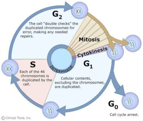
The first thing to understand about cell replication is the larger cycle the cell goes through in order to replicate. Fig 1 is an illustration of the cell cycle, cells usually perform most function during interphase. Within interphase is also when most normal cellular function is carried out. In the first phase of interphase (G1) the cell duplicates most of its contents aside from its genetic material. Within this process there are also a variety of checkpoints, points when the cell checks for whether it is capable of replicating. A well known checkpoint is the restriction checkpoint also known as the eG1 checkpoint. The restriction checkpoint is when the cell checks for most of the factors needed for replication, such as DNA damage, sufficient nutrients, space, etc. If the cell does not pass the checkpoint it will pause to fix the damage or wait for conditions to suit and may even trigger processes to kill the cell, a cell regulating its own death is known as apoptosis. In S phase the cell replicates its genetic material, and in G2 it checks again for DNA damage before performing mitosis which is covered later in this section.
Another thing to mention is how cells are moved through the cell cycle and how this process is triggered. All healthy cells rely on a variety of different signals to tell them when to replicate, for some cells this could include hormones or cytokines, all of these signals are grouped into the term “growth factors”. These growth factors can be released in a variety of different events, platelets can release cytokines to promote regrowth and healing (such as when the skin is cut and skin cells around the area grow to fill the gap), various glands can produce hormones in response to age that promote growth in specific cell types (such as in puberty when female breast tissue develops) and much more. Growth factors often cause a production of cyclins, there are a variety of different cyclins that activate different cyclin dependent kinases. Cyclin dependent kinases (CDKs) are present in cells for the majority of the cell cycle but are only active once cyclins are introduced in interphase. CDKs catalyze a variety of cellular processes that move the cell through the different phases and checkpoints of interphase. To summarize, chemical signals can trigger the production of cyclins, when cyclins are produced they activate CDKs and CDKs then help other proteins move through the steps needed to replicate.
More on the specifics of the checkpoints mentioned here:https://pmc.ncbi.nlm.nih.gov/articles/PMC4990352/.
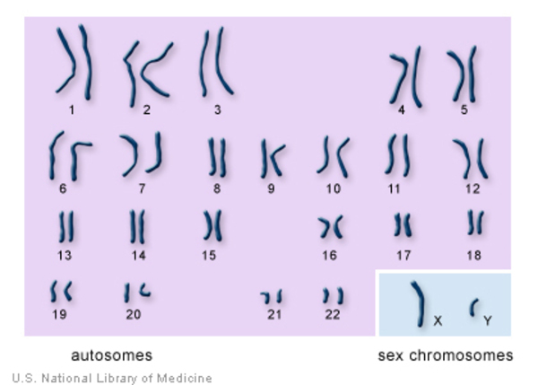
Before going into mitosis, and to a lesser extent meiosis, it is important to understand chromosomes. Chromosomes are bundles of DNA. A healthy human’s entire DNA sequence is made of 23 pairs of chromosomes, forming 46 in total. Each pair of chromosomes have one from each parent. Sex chromosomes carry information regarding the biological sex of the person, inheriting the chromosomes named Y and X causes a person to develop into a male, while 2 X chromosomes causes the person to develop into a female.
It is important to note that like many things in biology it is not universally agreed upon which way to organise the process of mitosis. There is an explanation of some of the variations at the end of the segment so the reader is able to understand how other sources may classify them slightly differently.
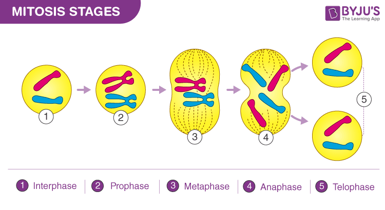
Fig 4 : A diagram showing the stages of mitosis.
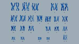
- Interphase is marked as the phase between replication, generally cells spend most of their life in interphase. Interphase can be separated into further phases once again. G1 and G2 are often referred to as the phases before DNA replication (G1) and after DNA replication (G2). More on the specifics of DNA replication here. S phase is the phase where DNA is replicated, this can get confusing because after replication there are now 2 copies of each pair of chromosomes, these 2 copies can remain joined for some phases of replication forming the classic X shape. Each half of these copied chromosomes are called chromatids (although the term chromatid is usually only used when discussing replication, after replication each separate chromatid is often referred to as a chromosome), 2 chromatids that once formed a chromosome together can be referred to as sister chromatids. Fig 2 and Fig 3 show chromosomes organised in both single strand and double stranded forms respectively. A cell spends most of its life in interphase so most regular cellular function happens in interphase.
- In prophase the DNA has officially replicated and is now organised and clear within the nucleus as the nuclear membrane dissolves. Mitotic spindles also begin to develop in prophase.
- In metaphase mitotic spindles attach to the kinetochore of each chromosome and apply equal force in each direction bringing the chromosomes into the centre of the cell.
- In anaphase the sister chromatids are separated at the centromere. They are then pulled to each pole of the cell.
- In telophase new nuclear membranes form around the two clusters of chromatids. Some consider cytokinesis a separate phase. Regardless of the characterization, in cytokinesis the space between the 2 nuclear membranes constricts, eventually forming 2 separating membranes.
Some people may say there are 4 stages to mitosis as they do not count interphase as part of the process(1,2,3), they do not disagree that this happens, they simply do not consider it a part of the process of mitosis. Others may say there are 6 or even 7 stages by breaking down prophase into prometaphase and prophase(2), and/or telophase into telophase and cytokinesis(2,3).
This process is important to understand as it highlights not only the mechanism cancer cells have disrupted, but also how they pass on these mutations. In interphase these cells make an exact copy of the DNA, as this is an exact copy it contains the exact same information and mutations the parent had. More on DNA replication here.
Meiosis is a slightly more complex process but will be covered in less detail as it is only mentioned for context.
Meiosis is the process specialised cells undergo to create cells used for sexual reproduction. These cells are called gametes or sex cells. The way gametes are produced leaves each gamete with only half of the required chromosomes(haploid), then when they combine with other gametes they can form a full set of chromosomes, leading to a diploid embryo (in humans specifically a sperm and ovum must meet).
There is also a process these cells undergo called crossing over. Simply put, each of the pairs of chromosomes pass small sections of their DNA to each other, thus promoting genetic diversity and ensuring that part of each chromosome is passed on to the next generation.
More information can be found here: https://byjus.com/biology/mitosis/
| 1. | Khan Academy. Phases of Mitosis. 2015. Accessed 6 August 2026. https://www.khanacademy.org/science/ap-biology/cell-communication-and-cell-cycle/cell-cycle/a/phases-of-mitosis |
| 2. | University of Leicester. The Cell Cycle, Mitosis and Meiosis for Higher Education | Virtual Genetics Education Centre | University of Leicester. 2023. Accessed 6 August 2026. https://le.ac.uk/vgec/topics/cell-cycle/the-cell-cycle-higher-education |
| 3. | Rehman I, Basit H, Malik A, Simpson B. Genetics, Mitosis. StatPearls Publishing; 2023. Accessed 6 August 2026. https://www.ncbi.nlm.nih.gov/books/NBK482449/ |