
DNA is arguably the most complex molecule in the human body, but also the most important to understand in the context of cancer. DNA has been used by pop culture since it was discovered, which has left a lot of people with half formed and slightly distorted ideas of what this elusive molecule is. Simply put, it is the fundamental recipe for all living things on earth.
Fig 1 Is a drawing of the molecule using the Watson and Crick model (simply named after the researchers who developed the model, it is the modern day accepted structure of the molecule. The image most people are familiar with is the twisted shape or double helix structure on the right of Fig 1. Although this is the natural state of DNA it brings unnecessary confusion to the simple structure. Think of DNA as a ladder or train tracks, the ladder is twisted into a helix in its natural state, however thinking of it untwisted and straight makes it much simpler.
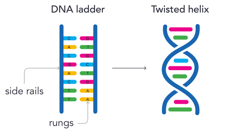
Fig 1 Watson and Crick model of DNA.
The “side rails” are alternating sugar and phosphate molecules, this is usually referred to as the phosphate backbone in scientific literature. However the most important part of the structure is the centre where you find the “rungs” of the ladder. These are nitrogenous bases, there are 4 nitrogenous bases: Adenine (A) Thymine (T) Guanine (G) Cytosine (C) . Each of these nitrogenous bases are fixed on one end to the sugar part of the sugar phosphate backbone, and on the other to another nitrogenous base. Each nitrogenous base can only connect with 1 other nitrogenous base, A-T and G-C. These 2 pairs are known as the base pairs. Fig 2 is a line structure diagram of a sequence of DNA. If Fig 2 does not make sense a tutorial on how to read chemical diagrams can be found here.
Note: A single segment of DNA (1 nitrogenous base, 1 sugar, and 1 phosphate) is known as a nucleotide.
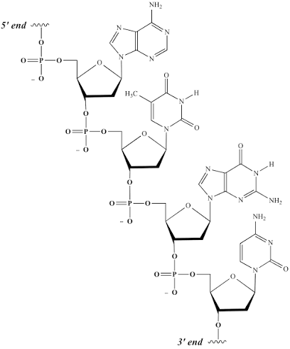
Fig 2 A line diagram of an example DNA sequence.
DNA is arranged into chromosomes during mitosis, and although DNA spends the majority of its time loose within the nucleus chromosomes are often mentioned when discussing DNA as the structures allow for easier categorisation of parts of DNA. Chromosomes are simply condensed DNA. DNA is wound around proteins called histones and condensed into the groups known as chromosomes. Chromosomes could be compared with balls of yarn, simply winding DNA up so as to keep different parts separate and organised. Refer to Fig 3 for a visualisation of chromosomes vs DNA.
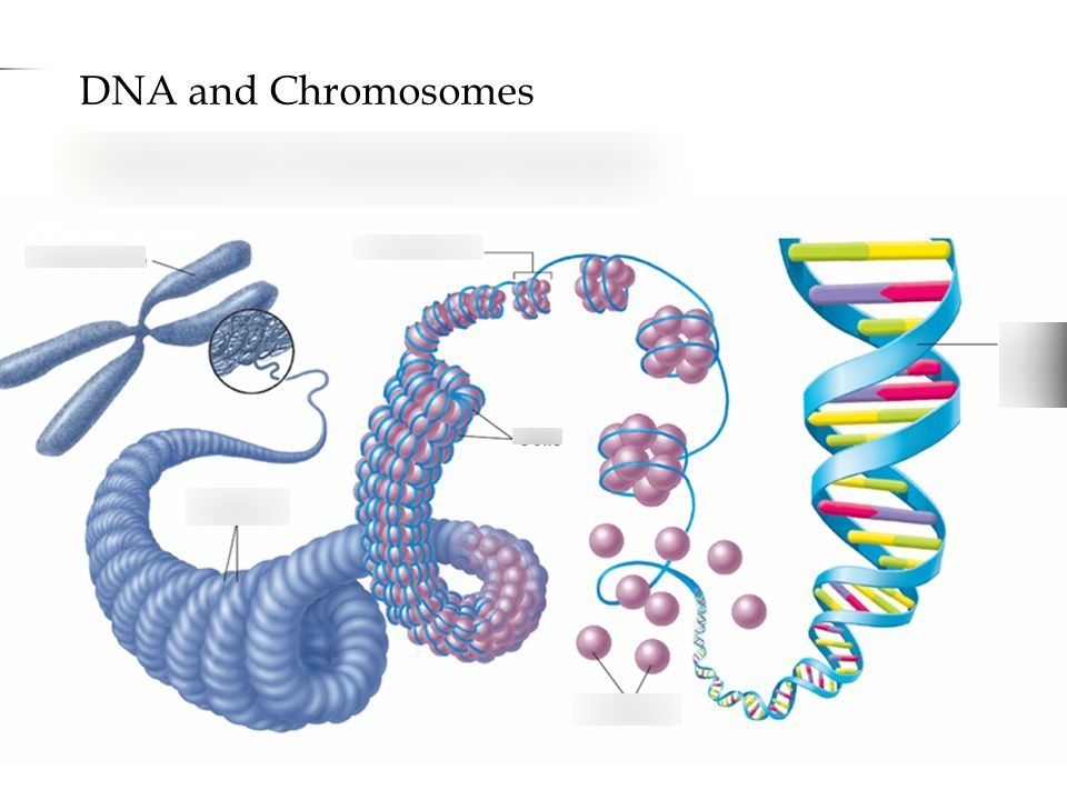
Fig 3 An illustration of how DNA is wound into chromosomes.
In practice each of the nitrogenous bases are a letter in the 4 letter alphabet of DNA. If you are familiar with morse code or binary you will already understand how alphabets can be made of 2 letters (dashes and dots, 0’s and 1’s). When coding for proteins, these nitrogenous bases are read in codons, which are sequences of 3. Each codon codes for a specific amino acid, these amino acids are then attached in the same order the codons are in. More on protein synthesis here.
Some people become caught up by the double stranded nature of the molecule, this is simply a redundancy that allows for easy replication and repair, since each nitrogenous base only pairs with one other, both sides have an opposite or mirrored version of the other strand. For example, adenine is only able to bind with thymine, therefore in any normal DNA molecule if there is an adenine base it will be connected to a thymine counterpart. If the adenine was then damaged, the cell could introduce a mix of free nitrogenous bases and only adenine could bind to the thymine and retake its correct spot.
When reading DNA proteins are able to determine which side to read off by the direction of the sugars. The sugar in DNA is not chemically symmetrical, however it does have a 5 carbon ring, each of the carbons within that carbon ring have been named 1′ through to 5′. Towards one side of the sugar is the 5’ end and towards the other is the 3’ end (pronounced 5 prime and 3 prime). 5’ and 3’ attach to phosphate groups while 1’ attaches to the nitrogenous base. Proteins to do with protein synthesis or DNA replication can often only perform their function in a specific direction meaning they must often orient themselves based upon this chemical asymmetry. Fig 2 has a fully drawn sequence of DNA if the reader would like to see the position of the carbon ring in relation to the entire DNA molecule.
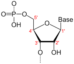
Fig 4: A line diagram of the chemical structure of the phosphate backbone of a DNA molecule. Note: The “Base” label in the diagram refers to where a nitrogenous base would normally be connected.
DNA replication is a conceptually simple process however depending on the detail it can get much more complex. Below is a simple understanding of the process that allows for visualization of the replication, after that is a more detailed explanation that includes names and terminology that will be useful if the reader intends to do further research. Some treatments and processes may function based on the intricacies of this process, so in depth understanding is recommended even if not necessary.
Simply put, the base pairs or “rungs” of the DNA are separated or “unzipped”. Then a variety of enzymes work together to keep the strands separate and call in more free nucleotides and essentially glue the new ones in place. These enzymes shouldn’t need to worry much about errors or misplacements because each nitrogenous base can only join with its corresponding pair making it only able to connect in the correct place.
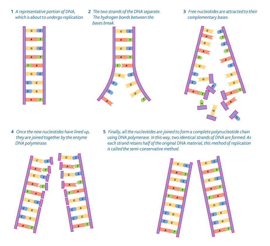
Fig 5: A simple graphic illustrating the base mechanisms for DNA replication.
The intricacies of this process have been thoroughly investigated. The first step of replication is for an enzyme known as helicase to break the hydrogen bonds connecting the nitrogenous bases to each other. Next an abnormal nucleotide known as RNA primer is added to one of the bases to form a starting place for the next protein, DNA polymerase III attaches to the strand and helps the new nucleotides to form hydrogen bonds with each other. This builds a second strand slowly following the helicase duplicating the entire strand.
Of course it could not be this simple as DNA polymerase III is only able to work from the 5’ to the 3’ end. Because the 2 strands are going opposite directions only 1 DNA polymerase can simply follow the helicase as it unzips (this is called the leading strand), the other must start from the helicase (the position of the helicase can sometimes be referred to as the replication fork), then work its way back to the start of the strand before jumping back to the helicase and working back down (this is called the lagging strand). This jumping back and forth means that all of the strand is not connected and is instead slightly fractured in places, these are known as Okazaki fragments (after the head researcher of the team that discovered them). Each starting place must have another RNA primer to facilitate the start of the Okazaki fragment. DNA polymerase I then goes back over and repairs damage and ensures there are no issues with the new strand while DNA ligase bonds the Okazaki fragments together. DNA polymerase I then can remove the primer. If the process is performed correctly there should now be 2 separate double stranded forms of DNA.
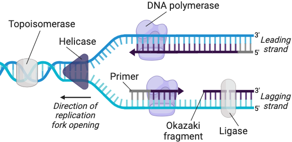
Fig 6: a more complete diagram of DNA replication.
More information can be found here: https://www.ncbi.nlm.nih.gov/books/NBK9940/