
A crucial aspect of cellular function to understand is protein synthesis, this is simply the process of making proteins. There are 3 main phases of protein synthesis: transcription, translation, and folding. Note: some institutions will refer to polypeptide synthesis rather than protein synthesis; this is the same concept but does not include the final step of folding.
Note: This section assumes the reader has read the section on DNA and Human cells.
During transcription a protein known as RNA polymerase will “unzip” a small portion of DNA, meaning it will break the hydrogen bonds holding the nitrogenous bases together. As covered in the DNA section each nitrogenous base can only bind with 1 other type, adenine to thymine and cytosine to guanine. This means that by unzipping the DNA, RNA polymerase can facilitate new RNA nucleotides binding with the original strand, resulting in a new strand of RNA that should be an inverse of the original.
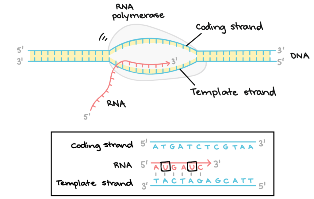
Fig 1: A graphic illustrating transcription.
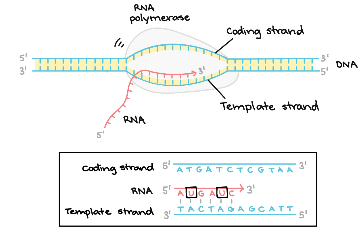
Note: RNA is slightly different to DNA because of small changes in chemical structure. The important change is that thymine is replaced by uracil, or in short form T is replaced by U. Fig 1 shows how RNA polymerase synthesizes RNA on the new strand, and below has a small diagram showing how this translates.
To follow the example sequence in fig.1, By allowing new nucleotides to bind with the DNA code
T A C T A G the cell creates a complementary strand of RNA that would follow the code
A U G A U C.
This RNA is then transported out of the nucleus and is known as mRNA or messenger RNA. Once the mRNA is outside of the nucleus the cell will then begin the next step of protein synthesis, translation. Ribosomes facilitate translation by clamping onto mRNA. Ribosomes have their own rRNA or ribosomal RNA, this means ribosomes are able to read key start and stop codons to ensure they are reading the correct area. Once the ribosome is clamped onto the mRNA the last type of RNA, tRNA or transfer RNA is called in. These proteins bind with specific amino acids that correspond to its specific anti-codon. Codons are the words of this biological language, in protein synthesis codons are 3 nitrogenous bases long and each of these combinations codes for an amino acid. Anti-codons are the inverse of the original codon, this means tRNA is able to bind to specific sets of nucleotides and therefore bind the amino acids into the correct place along the polypeptide chain. More information can be found here: https://www.ncbi.nlm.nih.gov/books/NBK558999/.
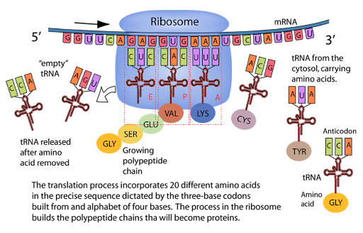
Fig 2: A graphic illustrating and explaining the entire process of translation.
Researchers have been able to identify the codes for each of the amino acids as shown in Fig 3. The 3 letter abbreviations next to the codons are abbreviations for amino acids, for example AAA and AAG will code for Lys or lysosome. UAA UAG and UGA code for the end of translation and are referred to as stop codons.
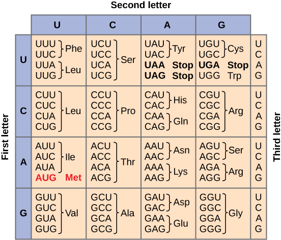
Fig 3: A table showing what amino acid each codon codes for.

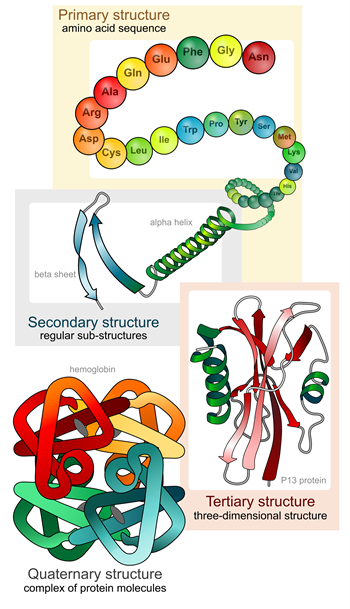
The final part of protein synthesis is folding. At the end of translation the cell is left with a chain of specific amino acids bound in a single file line, known as a polypeptide. This chain is then required to fold for the polypeptide to become a functional protein. The way this is done varies depending on the specific protein, but it should be noted that all of the information needed to fold into the correct form is contained within the structure itself. Meaning that although some molecules can help, and the environment it is in is important, none of that dictate how it folds. The polypeptide chain itself is the only thing that dictates its eventual shape which is sometimes referred to as its native fold.
There are also different levels of structure when referring to a protein. Primary structure is the basic sequence of amino acids, secondary structure is the physical structure the polypeptide chain forms, and tertiary structure is the folded form of the polypeptide. Some proteins also join together to form another structure referred to as a quaternary structure. (1)
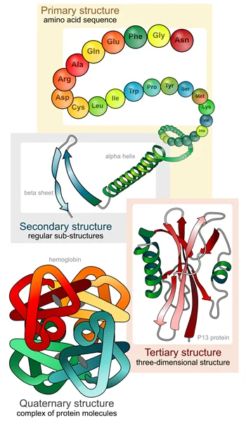
Fig 4: An illustration of each level of structure a protein can have.
Context: Like all things in biology, folding can go wrong. In most cases this causes the protein to simply be inactive, however in specific cases this can cause it to become misfolded in a way that makes it toxic. Diseases such as Creutzfeldt-Jakob disease, or the more commonly known version found in cattle: mad cows disease, is caused by a misfolded protein that misfolds other brain proteins as well. Similarly, clumps of abnormal proteins have been found in brains of people diagnosed with neurodegenerative diseases such as Parkinson’s or Alzheimer’s, although it is still unknown if this is a cause or symptom. (2)
Once a polypeptide is folded it is officially considered a protein.
| 1. | Everse S. Protein Folding. comis.med.uvm.edu. Published 2014. Accessed July 11, 2026. https://comis.med.uvm.edu/VIC/coursefiles/MD540/MD540-Protein_Organization_10400_574581210/Protein-org/Protein_Organization8.html |
| 2. | Disorders FNNS, Policy BHS, Medicine I. Protein Aggregation. National Academies Press (US); 2013. Accessed July 11, 2026. https://www.ncbi.nlm.nih.gov/books/NBK208522/ |